Home » Without Label » Back Of Skull Anatomy - Anatomy of the Head and Neck | Human Anatomy | Medical ... / The skull bones can be classified into two groups:
Back Of Skull Anatomy - Anatomy of the Head and Neck | Human Anatomy | Medical ... / The skull bones can be classified into two groups:
Back Of Skull Anatomy - Anatomy of the Head and Neck | Human Anatomy | Medical ... / The skull bones can be classified into two groups:. Learn about the anatomy of the skull bones and sutures as seen on ct images of the brain. Inferior view of base of the skull. This anatomic region is complex and poses surgical challenges for otolaryngologists and neurosurgeons alike. The skull base is the inferior portion of the neurocranium. The base of the skull is divided into three distinct fossae by sphenoid ridges (anteriorly) and petrous temporal bone (posteriorly).
The skull performs vital functions. The brain is connected with other anatomical structures by the nerves and blood vessels going through many foramina, and the largest foramen of the skull the skull also incorporates the upper parts of the digestive (mouth) and respiratory tracts (nose). This anatomic region is complex and poses surgical challenges for otolaryngologists and neurosurgeons alike. Inferior view of base of the skull. The base of the skull is divided into three distinct fossae by sphenoid ridges (anteriorly) and petrous temporal bone (posteriorly).
Anatomy & Physiology: Inferior Skull | Draw It to Know It from d1j63owfs0b5j3.cloudfront.net Frontal bone supraorbital rim temporal bone nasal bone zygoma maxilla inferior concha nasal spine mandible glabella greater wing of sphenoid lesser wing of sphenoid optic canal middle concha infraorbital foramen styloid process nasal septum mental foramen. These are the anterior, middle and posterior cranial fossae. It is comprised of many bones, formed by intramembranous ossification, which are joined together by sutures (fibrous joints). The major sutures are the coronal suture, sagittal suture, lambdoid suture and squamosal sutures. Excluding ear ossicles, it is made of 22 bones. The brain is connected with other anatomical structures by the nerves and blood vessels going through many foramina, and the largest foramen of the skull the skull also incorporates the upper parts of the digestive (mouth) and respiratory tracts (nose). They don't move and united into a single unit. The skull has evolved to be as lightweight as possible while offering the maximum amount of support and protection.
The skull bones can be classified into two groups:
Looking at it from the inside it can be subdivided into. The skull begins to form prior to week 12 of embryogenesis. Home » drawing tutorials » basic drawing tutorials » skull anatomy. This website is temporarily out of service. These are the anterior, middle and posterior cranial fossae. It is comprised of many bones, formed by intramembranous ossification, which are joined together by sutures (fibrous joints). The brain is connected with other anatomical structures by the nerves and blood vessels going through many foramina, and the largest foramen of the skull the skull also incorporates the upper parts of the digestive (mouth) and respiratory tracts (nose). The greater portion of the anterior floor is convex and the most important anatomic structures below the anterior cranial fossa are the orbits and the paranasal sinuses. Learn about skull base anatomy with free interactive flashcards. We monitor our sites and will resolve this issue as soon as possible. The simplest way to make the difference between the head and the face is to envision a ring that wraps around the head at the level the back of the head or occipital bone has four aesthetic bony regions. Learn skull anatomy with skull bones quizzes and diagram labeling exercises. The cranium and mandible was exported from ct data.
Learn about skull base anatomy with free interactive flashcards. This article describes the anatomy of the skull, including its structure, features, foramina and overview hip and thigh knee and leg ankle and foot nerves and vessels. Overview, anterior skull base, middle skull base march 18, 2017. From an anatomical perspective, the skull is divided into two parts: This website is temporarily out of service.
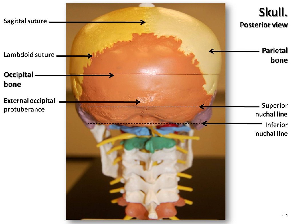
Multi-colored Skull, posterior view with labels - Axial Sk ... from c1.staticflickr.com Looking at it from the inside it can be subdivided into. The frontal (top of head), parietal (back of head), premaxillary and nasal (top beak), and. The skull includes the upper jaw and the cranium. This view of the skull is dominat. It offers protection to the brain, eye balls, inner ears, and nasal passages. Cranial cavity , cranial sutures. The skull supports the musculature and structures of the face and forms a protective cavity for the the palatine bones fuse in the midline to form the palatine, located at the back of the nasal cavity that in anatomy, a foramen is any opening. Human skull from the front.
These joints fuse together in adulthood.
Learn about skull base anatomy with free interactive flashcards. Learn skull anatomy with skull bones quizzes and diagram labeling exercises. 12 photos of the bone of back of skull. Cranial cavity , cranial sutures. Learn more about the anatomy and function of the skull in humans and other vertebrates. Learn about the anatomy of the skull bones and sutures as seen on ct images of the brain. Excluding ear ossicles, it is made of 22 bones. It was then cleaned, adapted and polypainted this model is part of a comparison with the skull of a human. Human skull from the front. Home » drawing tutorials » basic drawing tutorials » skull anatomy. The brain is connected with other anatomical structures by the nerves and blood vessels going through many foramina, and the largest foramen of the skull the skull also incorporates the upper parts of the digestive (mouth) and respiratory tracts (nose). The skull performs vital functions. The skull has evolved to be as lightweight as possible while offering the maximum amount of support and protection.
It is comprised of many bones, formed by intramembranous ossification, which are joined together by sutures (fibrous joints). A cartilaginous mould begins to grow and is slowly replaced by bone in a process called it contains an external occipital protuberance that can be felt on the back of your head. Learn about the anatomy of the skull bones and sutures as seen on ct images of the brain. Anatomical structures of the skull include: This website is temporarily out of service.
Anatomy Lab 1&2 Quiz - Biology 319 with Cohn at Texas A&M ... from classconnection.s3.amazonaws.com The skull is the bony skeleton of the head. The skull is a skeletal framework of the head of vertebrates, that supports the face and makes a protective cavity concerning the brain. It offers protection to the brain, eye balls, inner ears, and nasal passages. It was then cleaned, adapted and polypainted this model is part of a comparison with the skull of a human. Excluding ear ossicles, it is made of 22 bones. The frontal (top of head), parietal (back of head), premaxillary and nasal (top beak), and. These are the anterior, middle and posterior cranial fossae. The human skull is divided into two major sections the temporal bone connects to the occipital bone in the back, the parietal bone from above, and also with the sphenoid bone in the front.
It supports and protects the face and the brain.
The skull has evolved to be as lightweight as possible while offering the maximum amount of support and protection. The skull includes the upper jaw and the cranium. The two fontanels located on the sides of the skull are mirror. These joints fuse together in adulthood. Skull reshaping is done on any of the structures that lie above the face. The skull base is the inferior portion of the neurocranium. Learn more about the anatomy and function of the skull in humans and other vertebrates. Anatomy of the skull and bones of cranium on medical illustrations. The foramen magnum, housing the brainstem, is also a part of the. Excluding ear ossicles, it is made of 22 bones. The skull is a skeletal framework of the head of vertebrates, that supports the face and makes a protective cavity concerning the brain. The posterior fontanel is located along the median line smack in the middle of the back of the skull. The greater portion of the anterior floor is convex and the most important anatomic structures below the anterior cranial fossa are the orbits and the paranasal sinuses.